Renáta Makó1, Árpád B. Palotás1, István E. Sajó2 1 University of Miskolc, Hungary2 Chemical Research Center of the Hungarian Academy of Sciences
INTRODUCTION
Air pollution is one of the most frequently mentioned environmental problems of the world. Especially, in addition to gaseous pollutants (CO2, NOx, etc.) solid particles, soots play a significant role in this as soot can absorb polycyclic aromatic hydrocarbons that are known to be very harmful to human health. The structure of substances built mostly from carbon atoms are extraordinarily varied from mostly random to a perfectly ordered graphitic structure, which was studied earlier [1]. Previous studies showed how HRTEM coupled with image analysis can help quantifying the structural differences [1] and that crystallite dimensions and interlayer spacing distribution of Diesel soot and carbon blacks vary by the degree of oxidation [2,3]. This paper presents the result of a study on soots. The methodology was based on X-Ray diffraction measurements and a search for parameters that exactly determine a given soot specimen. The findings complement earlier results.
EXAMINED SPECIMENS
In the course of the experiment ten different types of soots were examined. Five of them were carbon black made by definite technological processes (N-306, N-373, N-375, N-660, N-772), two soots were produced by burning candles (G-01, G-02), two soots were made from Diesel oil (D-01, D-02), and a graphitized soot made from oil, which was kept at 1600 °C, 1800 °C, 2000 °C for an hour (Grk) [Table 1]. The results were compared with a standard reference model of graphite with the following parameters.
Table 1 Parameters of reference model Graphite-6R
RESULTS
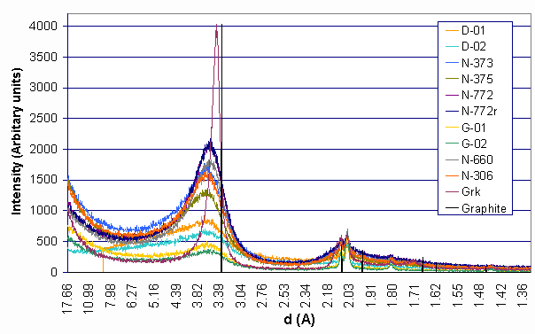
The specimens were examined with the help of a Philips PW 3710/PW1050 X-ray diffractometer (Cu Ka radiation, l = 0,1541862 nm, 40 kV, 35 mA, graphite monochromator, proportional counter). The results are summarised in Figure 1:
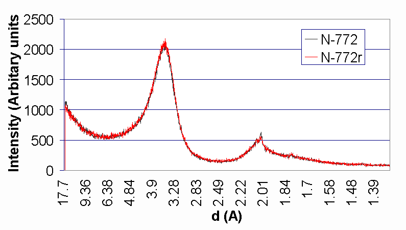
Figure 1 Intensity of carbon black and soot particulates Preliminary analysis of the diffraction patterns show similarities in the structure of the specimens to that of the graphite – as one can read in the literature on the subject. Structure of the carbon black specimens is more regular than that of soots, a reason of which can be probably the higher temperature of production. Intensity values of soots made from Diesel oil (D-01, D-02) and made from candle (G-01, G-02) are much lower than those of carbon black, because they have a looser structure and specimens with fewer mass can be compacted into the sample holder. The carbon black N-772 was reexamined to study how exactly the measurement process can be repeated. The yielding curve of the new sample from the same carbon black was designated as N-772r. Figure 2 shows the results of the two measurements. We established that the difference between the two curves is lower than 5 % around the first peaks of the curves. Figure 2 Intensity of carbon black N-772 and reproduction of its measure
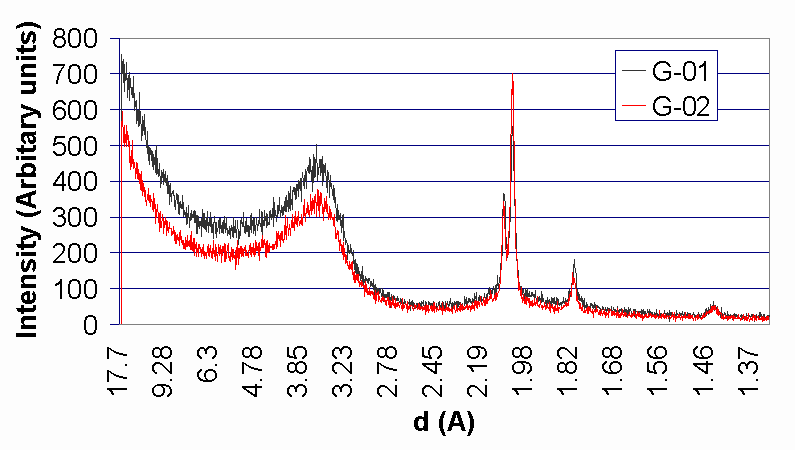
The results from the study of soots made from candle (G-01, G-02) and soots made from Diesel oil (D-01, D-02) can be seen on Figure 3 and Figure 4.
Figure 3 Profile of soots made from candle
Figure 4 Profile of soots made from Diesel oil
The shape of these curves are very similar indicating that there is a chance with the help of this measurement (and perhaps by comparing the results to those of other techniques) to distinguish certain types from each other.
CONCLUSION
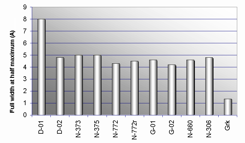
As a result of the study it can be stated that diffraction patterns of the examined carbon blacks, soots and graphite are similar. Further analysis and the determination of the full with at half maximum yielded the range of interplanar spacings characteristic of the specimens. Figure 5 shows the width values that range from 1,35 to 8 A. Under average interplanar distance we mean the one which belongs to the first peak of the curve. Displacements of these spacings happen mainly in direction of higher d values (looser structure). Full with at half maximum values of N-772 and N-772r are close to each other such as those of G-01 and G-02 according to our expectations, but the difference between values of D-01 and D-02 is considerably high (3,2 Å).
Figure 5 Full with at half maximum of each specimen
The average interplanar distance, which is one of the most important parameter of crystallised materials, is quite different (3,348-3,904Å) considering the values of Figure 6. It can be stated that the average interplanar distance value of Grk is the closest to that of Graphite and the difference of the values N-772 and N772r as well as those of D-01 and D-02 are fairly low (less than 0,035Å) as we expect, but it is quite high in the case of G-01 and G-02, which could be a result of a looser structure. The average distance in which samples can be considered crystalline state was 2 nm in most of the samples (except for soots made from Diesel oil, where it was closer to 1 nm, and graphitized soot, where it was 8 nm. It means that the specimens have a regular microstructure through 6 interplanar distance (in the case of graphitized soot it was 24).
Figure 6 Average interplanar distance of each specimen
REFERENCES
Copyright © 2000 |